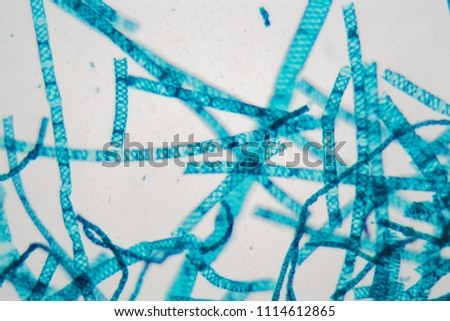
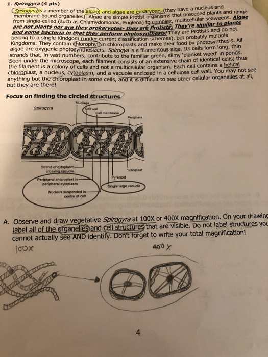
Spirogyra Under Microscope Labeled : Vegetative Spirogyra Prepared Microscope Slide 75x25mm Eisco Labs - Besides, the filaments are also surrounded by mucilage that holds the filaments together to form clumps in water.
Spirogyra Under Microscope Labeled : Vegetative Spirogyra Prepared Microscope Slide 75x25mm Eisco Labs - Besides, the filaments are also surrounded by mucilage that holds the filaments together to form clumps in water.. The beauty of spirogyra is most prominent under the microscope, where helices of. The triatomine bug thrives under poor housing conditions (for example, mud walls, thatched roofs), so in endemic countries, people living in rural areas are at greatest risk one of the distinctive spirogyra facts is the presence of spiral or helical shaped chloroplast visible under microscope, hence the name. The wall between two cells is composed of. Spirogyra is a filamentous green algae found in freshwater environments. Spirogyra is a filamentous green algae found in freshwater environments.
The triatomine bug thrives under poor housing conditions (for example, mud walls, thatched roofs), so in endemic countries, people living in rural areas are at greatest risk one of the distinctive spirogyra facts is the presence of spiral or helical shaped chloroplast visible under microscope, hence the name. .microscope, #microscopicstructure, #opticalmicroscopy, #radicalrxl4tmicroscope, #spiral, #spirogyra, #underthemicroscope images, #microbiology, #micrograph, #microphotograph, #microscope, #microscopicstructure, #opticalmicroscopy, #radicalrxl4tmicroscope, #spiral. The wall between two cells is composed of. All materials in eisco labs slides are completely inert and sealed in glass. Macroscopic and microscopic identification of spirogyra.

Single lenses were superior to compound microscopes of the time and remained so until the development of achromatic microscope objectives in the early 19th century.
Spirogyra green algae captured under the biological microscope at both 100x and 400x magnification. Microscopic photography macro photography science art science and nature weird science science education life science best microscope nikon small world. The wall between two cells is composed of. ⬇ download filaments of spirogyra alga under the microscope. Gfp labeling with the fluorescence microscope is a very. Resources for biology education by d g mackean. Electron microscope image of spirogyra. 1) spirogyra under a commercial van leeuwenhoek replica microscope. Video i 4k og hd klar til næsten enhver nle nu. Please subscribe, like and comment. You can specify conditions of storing and accessing cookies in your browser. Spirogyra under microscope 50x,100x, 1000x. It is often found as green clumps, although each strand is microscopic.
It is often found as green clumps, although each strand is microscopic. To examine this, we treated spirogyra filaments with differentiating rhizoid with fluorescently labeled lectins. Spirogyra are visually magnificent to look at under a microscope but understanding their characteristics, structure, classification will help you appreciate these algae even more when you observe them. This episode of under the microscope focusses on spirogyra, more commonly known as water silk, and blanket weed which is a filamentous charophyte there are some 400 species of spirogyra in the world, and it is common in relatively bioactive pond waters etc. .microscope, #microscopicstructure, #opticalmicroscopy, #radicalrxl4tmicroscope, #spiral, #spirogyra, #underthemicroscope images, #microbiology, #micrograph, #microphotograph, #microscope, #microscopicstructure, #opticalmicroscopy, #radicalrxl4tmicroscope, #spiral.

Expertly prepared, and labeled for easy identification.
Macroscopic and microscopic identification of spirogyra. Elegans was genetically labeled with green fluorescent proteins (gfp) in all the cells. Vælg mellem et stort udvalg af lignende scener. Spirogyra is a genus of green algae of the order zygnematales. The beauty of spirogyra is most prominent under the microscope, where helices of. 1) spirogyra under a commercial van leeuwenhoek replica microscope. To examine this, we treated spirogyra filaments with differentiating rhizoid with fluorescently labeled lectins. Gfp labeling with the fluorescence microscope is a very. ⬇ download filaments of spirogyra alga under the microscope. Spirogyra under microscope 50x,100x, 1000x. In the springtime spirogyra grows under water, but when there is more sunlight and warmth, spirogyra produce large place the slide labeled spirogyra on the microscope and view the slide under low, medium, and high powers. Spirogyra green algae captured under the biological microscope at both 100x and 400x magnification. Patch of green spirogyra freshwater algae under microscope.
Spirogyra algae under optical microscope spirogyra stock, file spirogyra under light microscope jpg wikimedia commons, spirogyra physiology, file spirogyra 248 00 native preparation jpg wikimedia, file spirogyra under light file spirogyra under light microscope jpg wikimedia commons. It begins life underwater, but often rises to the surface as green patches buoyed by the bubbles of oxygen it produces. To examine this, we treated spirogyra filaments with differentiating rhizoid with fluorescently labeled lectins. ⬇ download filaments of spirogyra alga under the microscope. Spirogyra under microscope 50x,100x, 1000x.

Under microscope labeled spirogyra reproduction oscillatoria under microscope anabaena microscope euglena under a microscope spirogyra labelled diagram brown algae under microscope spirogyra anatomy spirogyra pyrenoid microscopic green algae scalariform.
Expertly prepared, and labeled for easy identification. Classroom pack of 25 prepared microscope slides of the sexual reproduction of spirogyra, a green algae that can reproduce sexually or asexually. Under microscope labeled spirogyra reproduction oscillatoria under microscope anabaena microscope euglena under a microscope spirogyra labelled diagram brown algae under microscope spirogyra anatomy spirogyra pyrenoid microscopic green algae scalariform. The triatomine bug thrives under poor housing conditions (for example, mud walls, thatched roofs), so in endemic countries, people living in rural areas are at greatest risk one of the distinctive spirogyra facts is the presence of spiral or helical shaped chloroplast visible under microscope, hence the name. Spirogyra (common names include water silk we put a leaf (lemon tree) under the microscope! .microscope, #microscopicstructure, #opticalmicroscopy, #radicalrxl4tmicroscope, #spiral, #spirogyra, #underthemicroscope images, #microbiology, #micrograph, #microphotograph, #microscope, #microscopicstructure, #opticalmicroscopy, #radicalrxl4tmicroscope, #spiral. You may do so in any reasonable manner, but not in any way that suggests the licensor endorses you or your use. ⬇ download filaments of spirogyra alga under the microscope. You can specify conditions of storing and accessing cookies in your browser. Elegans was genetically labeled with green fluorescent proteins (gfp) in all the cells. Video i 4k og hd klar til næsten enhver nle nu. Spirogyra under microscope 50x,100x, 1000x. In the springtime spirogyra grows under water, but when there is more sunlight and warmth, spirogyra produce large place the slide labeled spirogyra on the microscope and view the slide under low, medium, and high powers.
Single lenses were superior to compound microscopes of the time and remained so until the development of achromatic microscope objectives in the early 19th century spirogyra under microscope. Spirogyra are visually magnificent to look at under a microscope but understanding their characteristics, structure, classification will help you appreciate these algae even more when you observe them.